Modern ophthalmology – individual & personal

At Augenzentrum Bachenbülach, we provide comprehensive ophthalmic preventive, diagnostic and assessment examinations for adults and children at the highest medical standard — guided by expertise, diligence, cost-effectiveness and clinical relevance.

Our services

Preventive check-ups for adults

From the age of 40, we recommend regular eye check-ups.
Preventive check-ups for children

From the age of 3 — or earlier in case of symptoms or a family history — we recommend an ophthalmic examination.

Vision test / precise refraction

If your vision is impaired, we determine the best possible correction values.

Emergency treatment

What to do in an eye emergency? When it matters most, we are here for you!

Aeromedical fitness assessment

Do you need a fitness assessment as a pilot, air traffic controller or cabin crew member? You've come to the right place!

Orthoptics

We provide thorough diagnostics and targeted treatment for squinting and other disorders of visual function.

Driving fitness test

We provide a professional assessment of your fitness to drive for the Road Traffic Office.

Pre- and post-operative care for eye surgery

If surgery is required, we advise you on your options!

Second opinions

Unsure about a diagnosis or treatment plan? Get an independent second opinion!

Retina consultations

We rely on state-of-the-art diagnostic methods to detect retinal diseases early and reliably.

Glaucoma consultations

Detect early, treat precisely, preserve eyesight! We provide thorough diagnostics and individually tailored glaucoma therapy.

Ophthalmic examination at the slit lamp

Preventive check-ups for adults

Regular preventive check-ups with an ophthalmologist help detect eye diseases early and avoid permanent damage. The eye is a complex organ that can be affected by a wide range of diseases and age-related changes. Many of these changes progress gradually and are not noticeable in their early stages, which further underlines the importance of regular preventive examinations.

When changes are detected early, they can usually be treated effectively.

We recommend the following examination intervals:

  • Age 15–40:every 5–10 years
  • Age 40–65:every 2–3 years
  • From age 65:every 1–2 years

In the case of a family history or systemic conditions such as diabetes, hypertension or autoimmune diseases, earlier and more frequent eye examinations are essential for the long-term preservation of your eyesight.

Particular attention is given to the early detection of glaucoma, with increasing risk from the age of 40, and age-related macular degeneration, which occurs more frequently from the age of 60.

Both conditions often develop unnoticed and, if left untreated, can permanently damage vision and significantly impair visual acuity.

Early detection is crucial!

Using state-of-the-art diagnostics, we quickly determine whether action is needed and plan the appropriate treatment together with you.

Child undergoing a vision test

Preventive examinations for children

Shortly after birth, infants begin to perceive their surroundings visually — at first only in a blurred way. Through daily stimulation and practice, eyesight develops step by step. Full visual acuity is typically reached between the ages of 6 and 8 and stabilises further from around age 10–12. Early ophthalmic preventive examinations are therefore important, even in children without apparent symptoms, in order to detect and treat potential vision problems in good time.

Amblyopia (lazy eye) — early detection is crucial

The first four years of life are decisive for visual development. During this sensitive period, even minor disturbances can prevent eyesight from developing fully.

If left untreated, this can lead to permanent amblyopia (lazy eye) — even in anatomically healthy eyes. Common causes include squinting, refractive errors, unequal visual acuity between the eyes (anisometropia), or congenital changes. Regular preventive examinations allow for early diagnosis and help prevent lasting visual impairment.

Children's glasses — optimal support from the start

If your child is found to have a refractive error, well-fitted children's glasses make an important contribution to healthy visual development. As children grow and their eyes continue to change, we regularly review and adjust their eyewear.

What matters most with children's glasses:

  • Lightweight, well-fitting frames
  • Flexible, padded temples and soft nose pads
  • Secure fit behind the ears
  • Lenses correctly positioned at eye height

What matters most with children's glasses:

Squinting — early detection and targeted treatment

Squinting is one of the most common causes of vision problems in childhood. At our practice, your child's eye alignment is examined by a specially trained orthoptist.

Orthoptists specialise in diagnosing and treating squint disorders, amblyopia, and disorders of binocular vision.

Depending on the findings, simple measures such as glasses or temporary patching of one eye can already be effective. If needed, we will advise you comprehensively on further treatment options.

Myopia control — effectively slowing short-sightedness

An increasing number of children are becoming short-sighted. The progression of myopia in childhood can now be actively influenced. We offer modern treatment methods such as special spectacle lenses (DIMS technology) as well as low-dose atropine eye drops.

We also regularly monitor eye growth by precisely measuring axial eye length, allowing us to assess exactly how quickly the eye is growing and whether myopia is progressing. This enables objective monitoring of myopia-control measures and individually tailored treatment.

Our goal is to slow excessive eye growth at an early stage and reduce long-term vision problems.

Eye test with a refraction device for glasses prescription

Vision test / precise refraction

Visual acuity can be reduced not only by refractive errors or issues with the cornea and lens, but also by retinal disease. This is why this examination is particularly important. During the vision test, we assess your current visual acuity without glasses or contact lenses.

We measure visual acuity at distance (from 5 m) and at near range (approx. 40 cm), with and without glasses or contact lenses as applicable, using standardised optotypes.

Using state-of-the-art measurement technology, including an automatic refractometer, we precisely determine the eye's refractive power — from short- and long-sightedness to astigmatism. We then verify the tolerability of the automatically measured values and, if required, the necessary correction for presbyopia.

On this basis you receive an individual spectacle prescription, which your optician can use to prepare eyewear perfectly tailored to your needs.

Eye emergency

Emergency care

In acute situations, we act quickly, carefully and with great empathy — so that you feel well cared for and your eyesight is protected as effectively as possible.

Common ophthalmic emergencies include:

  • Eye injuries (e.g. from foreign bodies or trauma)
  • Red eyes (particularly in contact lens wearers)
  • Visual disturbances (e.g. flashes, dark spots or soot-like specks)
  • Sudden loss or reduction of vision
  • Sudden double vision

During our daytime office hours, you can reach us by phone at 044 798 18 69.

We do everything possible to offer you an appointment as quickly as possible and to see you promptly at our practice.

Outside our opening hours, please contact the Doctors' Hotline at 0800 33 66 55.

You will be connected directly to the ophthalmologist currently on duty in the Zürcher Unterland/Winterthur region, who will provide competent support.

Aviation medical eye examination

Aeromedical assessments and flight safety

Good, healthy vision is essential in aviation. For aspiring and active pilots, air traffic controllers, private pilots and cabin crew, we carry out specialised aeromedical eye examinations. These examinations meet the requirements of the European Union (EASA) and the Federal Office of Civil Aviation (FOCA).

Professional pilots and air traffic controllers

Professional pilots and air traffic controllers must regularly demonstrate their fitness to fly in order to meet the high demands of flight operations. Examinations are based on Europe-wide regulations (EASA) and assess all required visual functions.

Private pilots

Private pilots must also have their fitness to fly regularly confirmed by a physician. The requirements are less strict than for professional pilots. If the visual function limits are not met during the examination by the Aeromedical Examiner (AME), a more extensive ophthalmic examination is indicated, which can be carried out by an ophthalmologist certified by FOCA.

Dr. Oroumchi is a recognised expert in aeromedical eye examinations for the Federal Office of Civil Aviation (FOCA) and is available to the medical advisors of Swiss Medical Services, the FOCA and the Aeromedical Institute (FAI) for questions regarding fitness to fly. Thanks to her thorough expertise and many years of experience, she ensures a reliable, standards-compliant assessment of visual performance in the aviation medicine context.

Book an appointment at our practice to have your aeromedical assessment carried out quickly and professionally. We ensure that all required examinations are completed smoothly and reliably.

Orthoptist testing a child's vision in front of the eye chart

Orthoptics

Orthoptics is dedicated to the assessment, prevention and treatment of squint disorders, amblyopia, neuro-ophthalmological conditions, and disorders of eye movement.

Our experienced orthoptist cares for patients of all ages with a high level of clinical precision and particular empathy.

A key focus is on the early examination and treatment of infants, pre-schoolers and school-age children. Especially with squint disorders, functional visual impairment and amblyopia, timely diagnosis is crucial for the successful development of vision.

Our goal is for your child to see clearly and sharply with both eyes after completing treatment — for a lifetime of optimal visual quality!

Orthoptic examination / vision therapy

A child's eyes go through an important developmental phase during the first years of life. It is precisely during this time that early detection and treatment of potential visual defects is crucial in order to avoid permanent visual impairment (amblyopia).

At our paediatric consultation, we take care of your children's eye health with empathy and professionalism.

Orthoptists are specially trained professionals for the examination and treatment of vision problems such as squinting, amblyopia, nystagmus and disorders of binocular coordination. A particular focus is placed on preventive examinations in children.

When should a child see an orthoptist?

  • Immediately, if abnormalities occur, e.g. nystagmus, a cloudy cornea, unusually bright pupils, strong light sensitivity, visible squinting or a drooping eyelid — or whenever you are unsure.
  • At 6–12 months, in cases of increased risk, such as premature birth, developmental delay, a family history of squinting, significant refractive errors, or known hereditary eye conditions.
  • At around 3 years of age, even in children without apparent symptoms, to detect hidden squinting or refractive errors early. These often go unnoticed and can lead to permanent visual impairment — particularly one-sided visual defects.

Squinting can also develop suddenly later in life. In many cases this is treatable, but it should always be assessed by an ophthalmologist.

An early ophthalmological-orthoptic examination at our practice offers the best conditions for detecting and successfully treating visual and squint disorders in good time.

View from a car onto the road

Driving fitness test

To ensure safety on the road, ophthalmic assessments are mandatory for certain categories of driving licence.

What is assessed in the driving fitness examination?

The ophthalmic driving fitness assessment determines whether your vision is sufficient for safe participation in road traffic. This includes testing:

  • Distance visual acuity, with and without glasses
  • Visual field
  • Eye alignment and eye movements
  • Vision in twilight and glare sensitivity* (where indicated)
  • Colour vision* (where indicated)
  • General state of health and the structures of the eyes

Examinations are carried out in accordance with the officially recognised requirements of the Swiss Traffic Admission Ordinance, ensuring a standardised assessment.

Eye surgery under the operating microscope

Pre- and post-operative care for eye surgery

If eye surgery is necessary, we refer you — independently and neutrally — to highly experienced eye surgeons using state-of-the-art techniques, for the best possible outcomes.

We accompany you from preparation through the procedure to aftercare, so that you feel safe and well cared for at every stage.

If you have any questions about the recommended eye surgery, please do not hesitate to contact us.

We are happy to support you with advice and guidance.

Medical consultation with a patient

Second opinion

There are often several ways to treat a condition. Especially with long-term medication or before surgery, the question frequently arises as to which approach is appropriate in an individual case.

We offer you a neutral assessment and comprehensive consultation.

We advise you competently, neutrally and in line with the latest developments in ophthalmology — so you can make the best decision for your eye health.

Ophthalmologist discussing a retina scan with a patient

Retina consultations

The retina forms the inner layer at the back of your eye and is a central component of your visual system.

High myopia, various conditions such as diabetes, hypertension or rheumatic disease, and certain medications can all lead to changes in the retina. We therefore work closely with your general practitioner to enable the best possible interdisciplinary care and treatment optimisation.

Retinal problems can sometimes occur suddenly: flashes, soot-like specks (“Russregen”) or floaters may indicate tears or detachments. Vascular occlusions or retinal inflammation can also occur.

The most common retinal diseases include diabetic retinopathy, macular degeneration, vascular occlusions and retinal detachment, among others.

A thorough retinal examination requires dilating the pupils with eye drops. This allows us to detect changes early — enabling timely intervention and the best possible preservation of your eyesight.

After pupil dilation, vision is reduced for several hours (approx. 3–5 hours), primarily at near range and to a lesser extent at distance, impairing reading ability and fitness to drive. If such an examination is planned for you, you should not drive yourself afterwards. Please ask if you are unsure whether pupil dilation will be necessary.

Ophthalmologist performing a slit lamp examination, glaucoma diagnostics

Glaucoma

Glaucoma refers to a group of eye diseases that can permanently damage the optic nerve. If left untreated, they lead to progressive visual field loss that increasingly restricts daily life. Glaucoma often remains unnoticed by those affected for a long time and, in the worst case, can lead to blindness! With gentle, precise examinations, we identify risks early and protect your eyesight for the long term.

Diagnosing glaucoma requires not only measuring intraocular pressure but also a biomicroscopic assessment of the optic nerve and, where abnormalities are found, further diagnostics such as visual field testing and optic nerve analysis using OCT (optical coherence tomography).

Once all findings have been reviewed together, the diagnosis can be confirmed and treatment initiated — in most cases with eye drops to lower intraocular pressure. Treatment success should be monitored every six months, or more frequently in certain cases.

Fast, non-contact, precise

Our practice is equipped with modern diagnostic devices. For further surgical procedures, we work closely with recognised specialists and renowned centres.
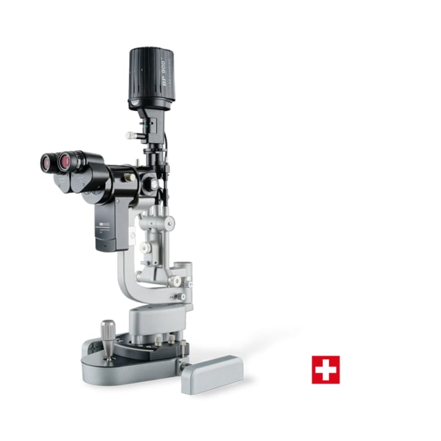
Slit lamp examination

Slit lamp examination

Slit lamp examination

The slit lamp allows a detailed examination of the anterior and posterior segments of the eye under high magnification. This enables precise assessment of the cornea, lens, conjunctiva and eyelids, and the early detection of the smallest changes.
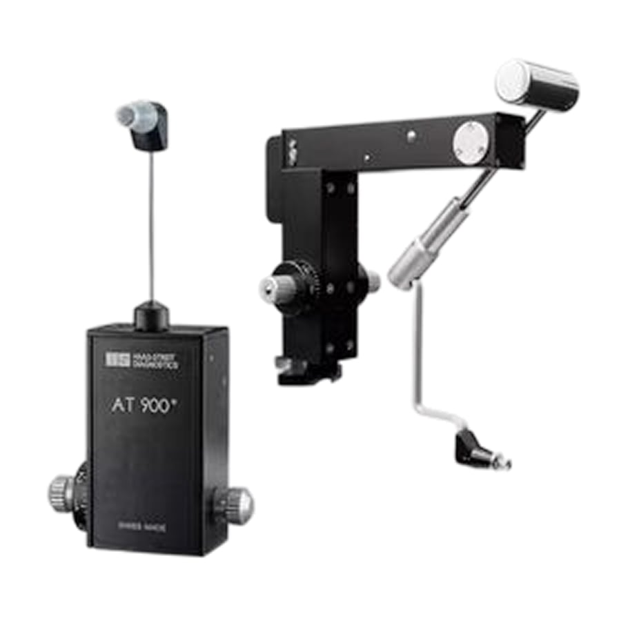
Intraocular pressure measurement / Goldmann tonometry

Intraocular pressure measurement / Goldmann tonometry

Intraocular pressure measurement / Goldmann tonometry

With this method, we measure the pressure inside your eye with particular precision. Elevated intraocular pressure can indicate glaucoma and should be detected early. The examination is painless, quick and highly reliable.
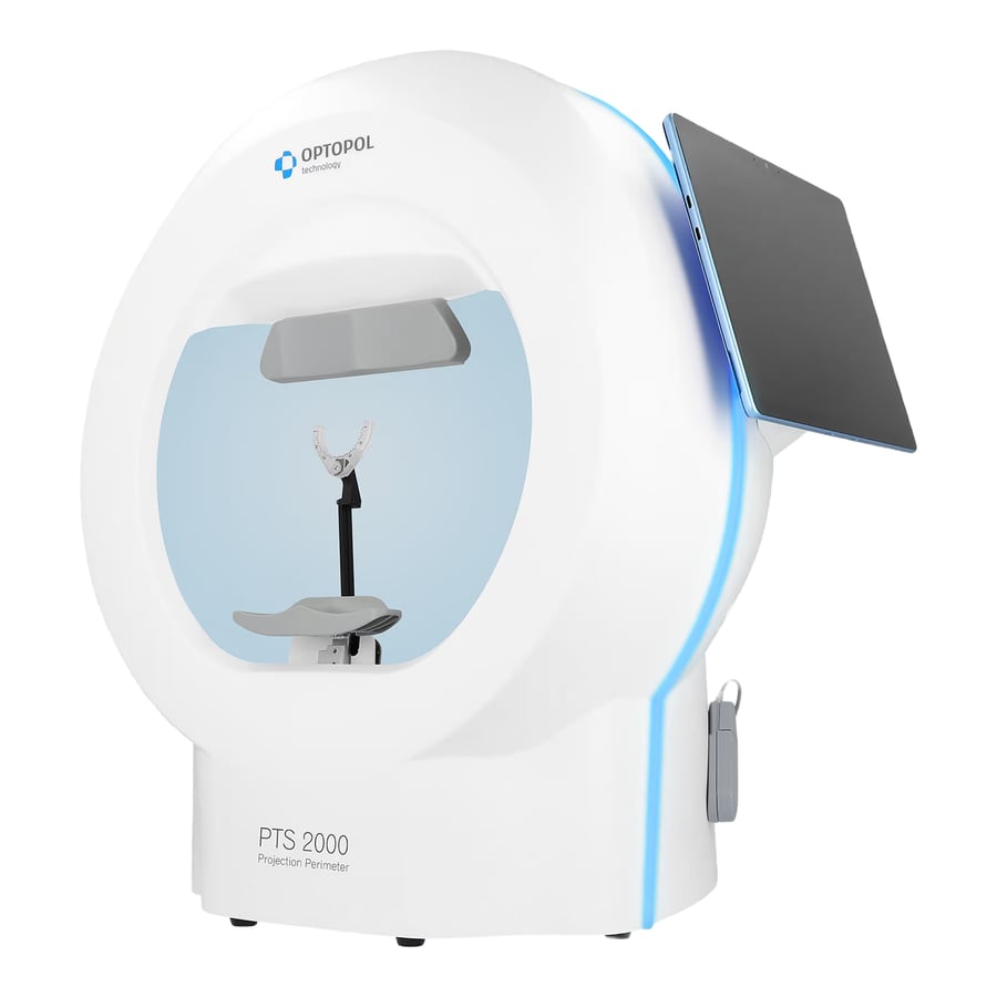
Visual field examination

Visual field examination

Visual field examination

Visual field testing assesses how well you see peripherally and centrally. It is particularly important for the early detection and monitoring of conditions such as glaucoma or certain neurological disorders.
Axial length measurement / biometry

Axial length measurement / biometry

Axial length measurement / biometry

With this device, we precisely measure the axial length of the eye. This measurement is especially important for short-sighted children, as the progression of myopia is directly linked to the growth in eye length.
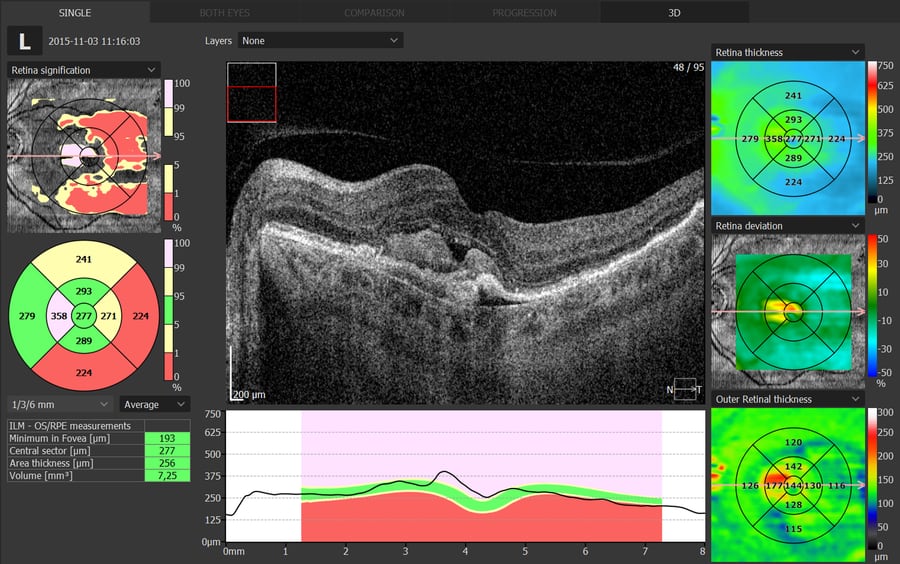
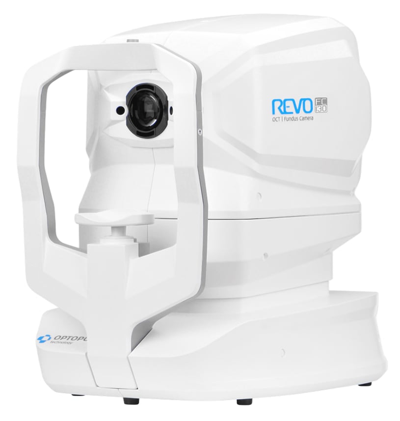
OCT cross-sectional imaging

OCT cross-sectional imaging

OCT cross-sectional imaging

OCT allows us to produce high-resolution cross-sectional images of the retina — non-invasively and painlessly. This enables early detection and precise monitoring of conditions such as macular degeneration or glaucoma.
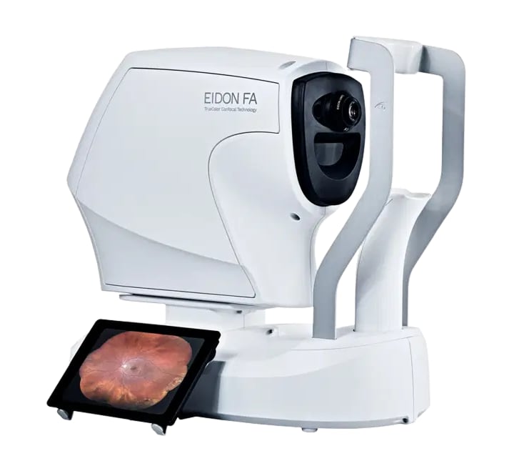
Retinal photography

Retinal photography

Retinal photography

The fundus camera produces colour images of the retina and optic nerve. These images serve as precise documentation and help reliably detect and monitor changes at the back of the eye.
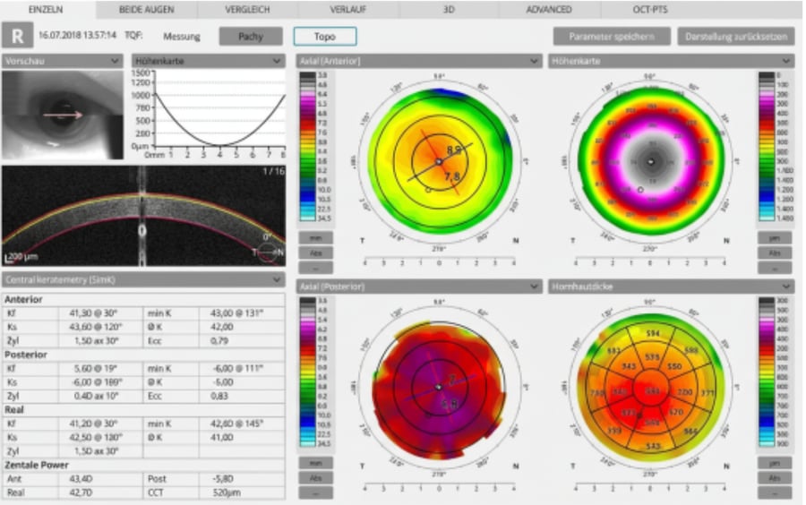
Corneal topography

Corneal topography

Corneal topography

The corneal topographer measures the surface of the cornea without contact and creates a highly precise, colour-coded map of its shape and curvature. This examination is particularly important for the early diagnosis of keratoconus — a condition in which the cornea gradually thins and bulges. The measurement is fast, painless and provides important information for diagnosis, monitoring and treatment planning.
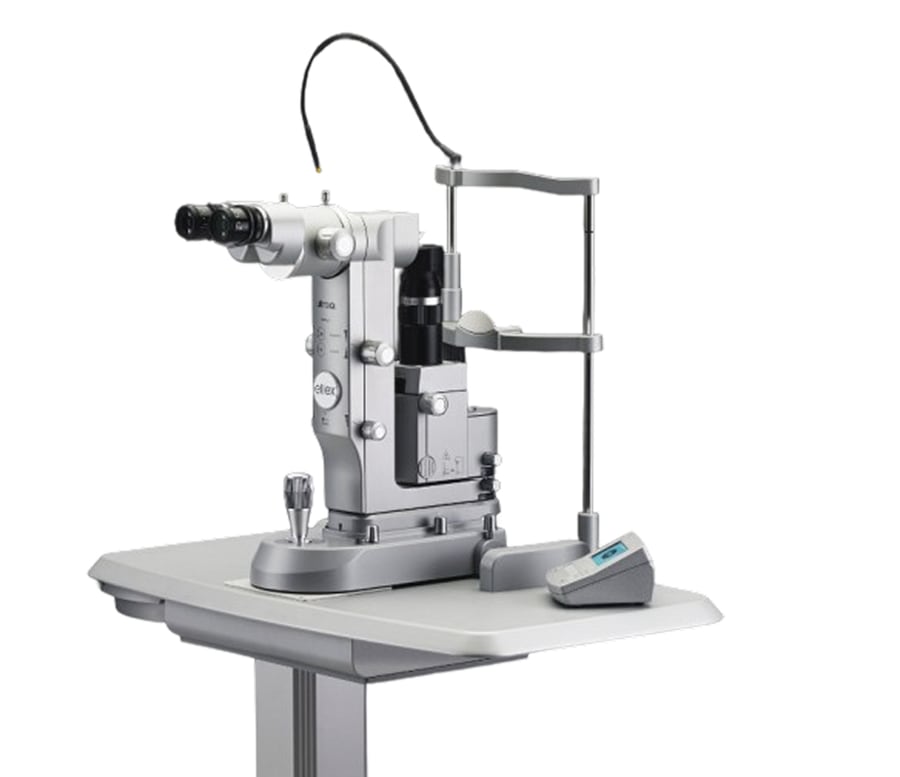
Laser treatments

Laser treatments

Laser treatments

Our laser allows certain eye conditions to be treated gently and precisely. The YAG laser, for example, is used to treat posterior capsule opacification, while SLT treatment can lower intraocular pressure in glaucoma — performed on an outpatient basis, with minimal discomfort and without any incision.
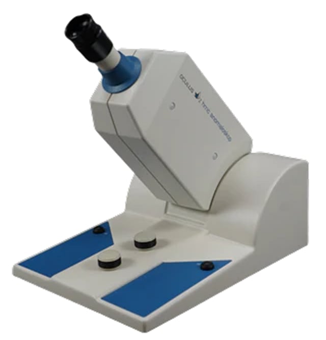
Colour vision test / anomaloscope

Colour vision test / anomaloscope

Colour vision test / anomaloscope

The anomaloscope allows a particularly precise assessment of your colour vision. The test shows whether colours — especially red and green tones — are perceived normally, or whether a colour vision deficiency is present. The test is painless, takes only a few minutes and delivers highly precise results.

Frequently asked questions – well informed before your visit

Answers to the most common questions about examinations, paediatric ophthalmology, eye diseases and our practice – medically sound and explained clearly.

  1. A regular check-up is worthwhile even without symptoms, because many eye diseases – including glaucoma and macular degeneration – remain symptom-free for a long time. As a guide, we recommend:

    • Age 15–40: every 5–10 years
    • Age 40–65: every 2–3 years
    • From age 65: every 1–2 years

    With risk factors – a family history (e.g. glaucoma), diabetes, hypertension, autoimmune disease, high myopia or certain medications – earlier and more frequent examinations are advisable. Whatever the interval, sudden changes in vision should be assessed promptly.

  2. The examination is tailored to your concern. It begins with a conversation about symptoms, previous conditions, medication and family history. Depending on the indication, the following steps then take place:

    • Visual acuity testing and determination of your spectacle prescription (refraction)
    • Assessment of the anterior segment of the eye at the slit lamp
    • Measurement of intraocular pressure (tonometry)
    • Examination of the back of the eye, including the retina and optic nerve head, often with dilated pupils
    • Where needed, additional diagnostics: optical coherence tomography (OCT), visual field testing, retinal photography or corneal topography

    The examinations are painless. Afterwards we explain the findings clearly and discuss the next steps.

  3. Only with a dilated pupil can the retina, macula and optic nerve head be assessed reliably out to the periphery – for example when retinal tears, diabetes, glaucoma or age-related macular degeneration are suspected. The eye drops (mydriatics) usually take effect within 15–30 minutes.

    Afterwards, near vision and glare sensitivity are impaired for roughly 3–5 hours. During this time, reading and driving are not safe. Please plan your journey home accordingly (accompanying person, public transport) and bring sunglasses. When booking, ask whether pupil dilation is planned for your examination.

    • Your current glasses and your contact lenses with their case
    • A list of your medication (including eye drops and over-the-counter products)
    • Previous findings and medical reports, if available
    • Your insurance card and, if applicable, your referral
    • Sunglasses for the journey home

    Please let us know if you wear contact lenses – depending on the examination, we may ask you to remove them beforehand.

  4. A sight test at the optician determines visual acuity and spectacle strength (refraction). An ophthalmological examination goes further: it assesses the health of every part of the eye – cornea, lens, intraocular pressure, retina and optic nerve – and detects diseases that initially cause no symptoms.

    If visual acuity remains reduced even with optimal glasses, a medical assessment is always indicated: the cause may be a disease of the retina, the optic nerve or the lens.

  5. For adults with stable prescriptions, a check every two years is usually sufficient. Come sooner if you notice increasingly blurred vision, headaches, or rapid fatigue when reading or working at a screen.

    From around the age of 40–45, the lens loses its ability to focus on near objects (presbyopia); reading or varifocal correction becomes necessary, and the prescription often changes gradually over the following years. Children and adolescents need more frequent checks because of growth – every 6–12 months, depending on the findings.

  6. We recommend a first preventive examination from around 3 years of age. It is advisable earlier with a family history (squint, amblyopia, high refractive error), after premature birth, with developmental delay, or when there are signs such as a visible squint, light sensitivity, a clouded cornea, an unusually pale pupil, trembling eyes or a drooping eyelid.

    The reason: vision develops during the first years of life. Disturbances in this sensitive period can lead to permanently reduced vision – the earlier they are detected, the better they can be treated.

  7. A squint is often visible but may also occur only intermittently. Signs include a tilted head posture, squeezing one eye shut in sunlight, frequent tripping, or holding books and screens very close.

    Amblyopia – reduced visual acuity resulting from disturbed development of the visual pathway although the eye itself is healthy – often goes unnoticed because the better eye compensates. Causes include a squint, differences in refractive error between the two eyes, or congenital changes. This is why preventive examinations matter. Treatment consists of glasses, temporary patching of the better eye and orthoptic training; our orthoptist supports your child throughout.

  8. Myopia progression can nowadays be slowed in a targeted way; there is no cure. Monitoring eye length is key: in myopia the eyeball grows too long. Regular axial length measurements let us assess how fast the eye is growing and whether a measure is working.

    Studies show effects for low-dose atropine eye drops and for special spectacle lenses (e.g. DIMS technology); spending enough time outdoors is regarded as effective prevention. Which measure is appropriate depends on age, baseline values and course. We summarise current studies in our Insights section.

  9. Glaucoma is a group of diseases in which the optic nerve is damaged step by step. The most important risk factor is raised intraocular pressure, although there are also courses with normal pressure. Further risk factors are older age (from about 40), a family history, high myopia and diabetes.

    Because the peripheral visual field is lost first and central vision is preserved for a long time, the disease is usually noticed late – lost optic nerve fibres do not regenerate. Diagnostics include measuring intraocular pressure, assessing the optic nerve head, visual field testing and OCT.

    Treatment consists of pressure-lowering eye drops, laser procedures (selective laser trabeculoplasty, SLT) or surgery. The aim is to preserve the remaining vision. Treatment is usually reviewed every six months.

  10. In a cataract, the natural lens of the eye becomes cloudy over the course of life. Typical signs are increasingly blurred, low-contrast vision, greater glare sensitivity (especially at night), muted, yellowish colours and frequently changing prescriptions.

    The clouding cannot be corrected with drops or glasses; only surgery is effective, in which the clouded lens is replaced by an artificial lens. The procedure is usually performed as an outpatient under local anaesthesia. The right time depends on how much it affects daily life (reading, driving, work) – not on a particular degree of “ripeness”.

    We clarify the indication, discuss lens options and look after you before and after the procedure. The surgery itself is performed by specialised surgeons with whom we cooperate.

  11. AMD affects the macula, the area of sharpest vision at the centre of the retina, and is among the most common causes of severe vision loss in older age. Warning signs are straight lines that appear distorted or wavy, a grey or dark spot in the centre, and increasing difficulty reading – you can check this yourself with an Amsler grid.

    A distinction is made between the dry form, which progresses slowly and is not curable (in certain stages, a nutritional supplement based on the AREDS2 formula, taken after medical advice, can slow progression), and the wet form with abnormal blood vessel growth. Untreated, it can lead to rapid vision loss and is now treated with medication injected into the vitreous (intravitreal anti-VEGF therapy). Risk factors are age, smoking and family history.

    If new distortions appear, an assessment with OCT should take place within a few days.

  12. Persistently raised blood sugar damages the small vessels of the retina (diabetic retinopathy) and can lead to macular oedema – an accumulation of fluid at the centre of the retina. Both cause no symptoms for a long time.

    This is why regular eye examinations, including examination of the back of the eye, are important for everyone with diabetes – usually once a year, more often if there are findings. The most effective prevention is good control of blood sugar, blood pressure and blood lipids. Depending on the stage, laser treatment, intravitreal medication or – rarely – surgery may be considered.

  13. Warning signs are sudden flashes of light, a swarm of black dots or threads (“floaters”), and a shadow or curtain moving into the visual field from the edge. Risk factors include high myopia, previous cataract surgery, eye injuries and a family history.

    This is an emergency: please have your eyes examined the same day. Retinal tears can be treated early with a laser; an already detached retina requires surgery – the earlier, the better the prognosis.

  14. Common eye emergencies include:

    • Eye injuries (e.g. from foreign bodies or trauma)
    • Red eyes, particularly in contact lens wearers
    • Visual disturbances such as flashes, dark spots or floaters
    • Sudden loss or reduction of vision
    • Sudden double vision

    First aid: For chemical burns (e.g. from cleaning products), rinse the eye immediately with plenty of clean water (at least 10–15 minutes, keeping the eyelids open) and then seek medical examination without delay. With foreign bodies or injuries, do not rub or remove anything yourself; cover the eye loosely. Contact lens wearers with a red, painful eye should remove their lenses and not reinsert them. During our consultation hours you can reach us on 044 798 18 69.

  15. Our consultation hours are Monday to Thursday from 8:30–13:00 and 14:00–17:00, Friday and Saturday by appointment. Outside these hours, please contact the Doctors' hotline on 0800 33 66 55: you will be connected directly to the ophthalmologist on duty in the Zürcher Unterland/Winterthur region.

    In life-threatening situations, call the emergency number 144.

  16. The easiest way is the online appointment booking (“Appointment” button in the header of this page). Alternatively, reach us by phone on 044 798 18 69, via WhatsApp (076 721 60 21), by email or through the request form. Please do not send health data or medical findings through these channels – we are happy to discuss details in person.

  17. Yes. Dr. Oroumchi has been a recognised ophthalmology expert for the Federal Office of Civil Aviation (FOCA) since 2019 and performs aeromedical eye examinations for professional and private pilots, air traffic controllers and cabin crew – in line with EASA and FOCA requirements.

    Please bring any existing documents as well as the requirements of your aeromedical examiner (AME) and mention the examination when booking.

  18. Yes. The practice is on the first floor of the Rietbach Center in Bachenbülach and is reached from the main entrance by stairs or lift – wheelchair- and stroller-friendly. By bus, take line 504 (Bramenstrasse stop, directly in front of the centre), lines 530/531 (Bachenbülach Alte Post) or line 501 (Bramenländli). Parking is available at the Rietbach Center (paid) and free of charge in the surrounding area.

  19. Cost coverage depends on the reason for the visit. Examinations for symptoms, or for the assessment and follow-up of a condition, are generally billed through compulsory basic health insurance. Purely preventive check-ups without symptoms, refraction for glasses, or certificates (e.g. aeromedical examinations, fitness to drive) may have to be paid for wholly or partly by yourself. Your insurance model and deductible are decisive.

    If in doubt, please clarify cost coverage with your health insurer beforehand; we are also happy to inform you before the examination.

The information on this page is for general orientation only and does not replace a personal medical examination or consultation.
Eye emergency

Emergency care

In acute situations, we act quickly, carefully and with great empathy — so that you feel well cared for and your eyesight is protected as effectively as possible.

Common ophthalmic emergencies include:

During our office hoursWe will offer you an appointment as quickly as possible.044 798 18 69 Outside opening hoursDoctors' hotline: connects you with the ophthalmologist on duty in the Zürcher Unterland / Winterthur region.0800 33 66 55

Prefer to call? 044 798 18 69